Electrical Signals Of The Brain To Control Movement
(Posted on Monday, September 25, 2023)

HANOVER, GERMANY: A mannequin is fitted with a prototype of a “mental typewriter”, which is an EEG (Electro Encephalogramme Measurement) Cap that processes electric impulses from the brain into commands for a computer at the Fraunhofer Institute for Computer Architecture and Software Technology stand during the 2006 CeBIT information and telecommunication technology fair 14 March 2006 in Hanover. he world’s biggest IT fair runs to 15 March, and some 6300 exhibitors of 71 countries will present their products. AFP PHOTO JOHN MACDOUGALL (Photo credit should read JOHN MACDOUGALL/AFP via Getty Images)
AFP VIA GETTY IMAGES
This was originally published on Forbes on 9/25/23.
This story is part of a series on the current progression in Regenerative Medicine. This piece discusses advances in brain-machine interfaces.
In 1999, I defined regenerative medicine as the collection of interventions that restore to normal function tissues and organs that have been damaged by disease, injured by trauma, or worn by time. I include a full spectrum of chemical, gene, and protein-based medicines, cell-based therapies, and biomechanical interventions that achieve that goal.
Brain-machine interfaces pose the opportunity to revolutionize modern regenerative medicine, but underlying research in brain signal generation, acquisition, and processing is still far from complete. One of the more difficult aspects of any brain-machine system is the conversion of brain waves from electrical signals in the brain to transferable data in the machine. While computer data is more or less a complex selection of ones and zeros, brain signals involve electrical and chemical relays. For brain-machine interfaces to progress, we must have an intimate understanding of brain signals and how they can be used in these systems.
A study in Frontiers by Dr. Usman Salahuddin and colleagues from the University of Connecticut discusses our current understanding of brain signal generation, acquisition, and processing. Here, I will use this article as a guide to comment on the state of brain signals regarding the future of brain-machine interfaces.
Before discussing how to acquire and decode brain signals, we must first understand the difference between brain signals and waves and how both impact brain-machine interface technologies.
The human brain is among the most complex organs on the planet. As Salahuddin notes, the brain contains three parts: the brainstem, limbic system, and cortex. The last of these controls processing, critical thinking, and analysis and is often the focal point for brain-machine interfaces.
Communication between the cortex and the rest of the body runs through the nervous system and, more acutely, the 100 billion neurons that run throughout the nervous system. Between these neurons are connection points, known as synapses, of which there are tens of trillions. Brain signals are the electrical impulses neurons use to communicate at the synapses.
Brain waves are similar but distinct from brain signals, as they are both related to electrical activity in the brain but differ in form. Brain waves are oscillating electrical voltages in the brain. Whereas a brain signal is a rapid milliseconds-long impulse, a brain wave may range up to several seconds in length.
There are four main types of brain waves, each associated with its own functions and state of being. These four are alpha, beta, delta, and theta waves.
The lesser-used brain waves in brain-machine interfaces are delta and theta waves. These are generated during sleep, yielding restorative effects and prompting creativity and visualization. It would be interesting to see these brain waves used in brain-machine interfaces for therapeutic use cases in the near future.
The more commonly used brain waves are alpha and beta waves. Alpha waves are associated with relaxation, focus, and concentration. These waves are commonly used in brain-machine interface activities requiring precision, such as controlling a cursor on a computer screen. Another example outside the brain-machine interface space is EndeavorOTC, a mobile game by Akili Interactive targeting children and adults with ADHD, improving focus and attention by improving alpha brain wave efficiency.
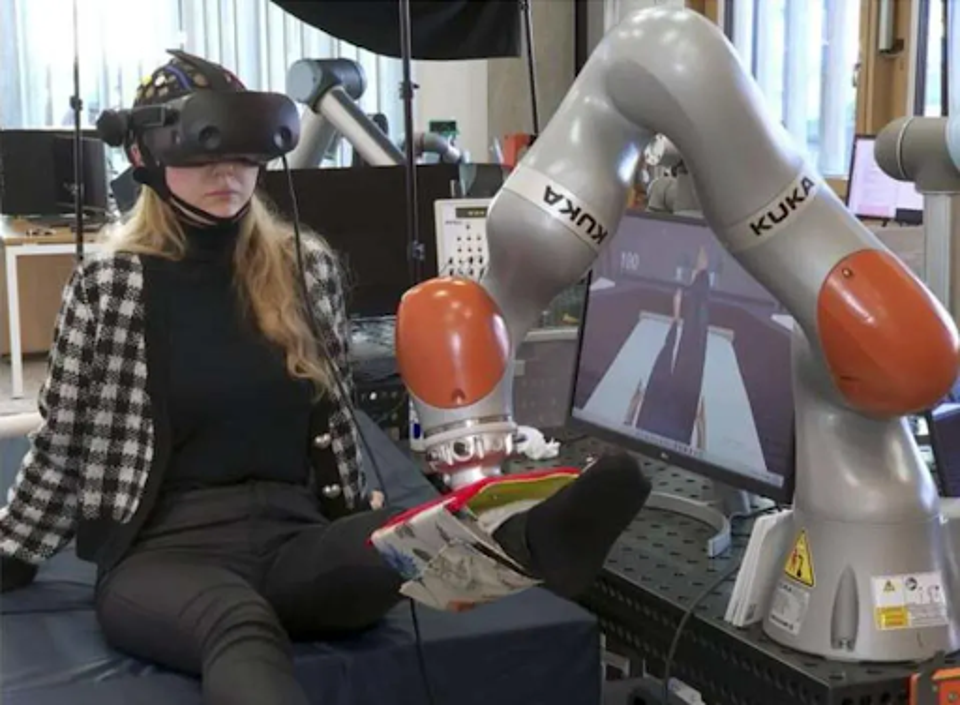
Beta waves are activated during baseline consciousness and are involved in standard mechanical functions like movement, exercise, and low-intensity mental activities. Beta waves are often used in prosthetic brain-machine interfaces, allowing users to move prosthetic limbs by thought. I have previously written on two such examples. One by researchers at the Skolkovo Institute of Science and Technology, where users activated a robotic limb with brain wave capturing software, and another by scientists at the University of Padova, where users could move their wheelchair using similar technology.
Brain activity is characterized by a combination of alpha, beta, delta, and theta waves, with one type of wave dominating during the person’s current state of being and consciousness. Efficiently capturing brain waves, in addition to brain signals, could yield a number of effective use cases for brain-machine interfaces in the coming years.
One way to measure both brain signals and brain waves is electroencephalography. This process measures electric signals generated by neurons within the brain, which are detected by placing electrodes on the scalp in the form of a headband or cap, such as that used in the Skolkovo and Padova examples. Brain-machine interfaces then take the recorded data from electroencephalography and use controlled algorithms to detect patterns to process the signals, yielding digital commands the interface can act on.
Scientists have developed a robotic prosthetic that offers new hope for patients suffering from traumatic spinal injuries.
SKOLKOVO INSTITUTE
Another method is that of magnetoencephalography. As can be deduced from its name, this method works similarly to electroencephalography but measures magnetic fields generated by the electrical activity of neurons as opposed to the electrical signals themselves. This process is also noninvasive, employing sensors outside the head. Whereas electroencephalography is most often used for communication and rehabilitation of people with motor impairment, magnetoencephalography is more therapeutic, used in preoperative and treatment planning for individuals with brain injury or disorders.
A third method to measure brain signals is intracortical recordings. Similar to electroencephalography, intracortical recordings record the electrical activity of neurons in the brain, but on an individual scale. This specificity is achieved by implanted electrodes in the brain parenchyma. This allows for high-resolution measurement of neural activity on a millisecond-by-millisecond basis. Intracortical recordings are often used in motor-based brain-machine interfaces, such as controllable prosthetic devices.
Implanted electrodes pose their own set of risks and challenges. These devices are comprised of many different materials typically found in computer equipment, such as platinum, iridium, and gold, but confined within scaffolding materials to enable long-term implant success such as silicon or ceramic. Metal electrodes implanted in the brain carry electrical current as the flow of ions, resulting in measured electrical signals.
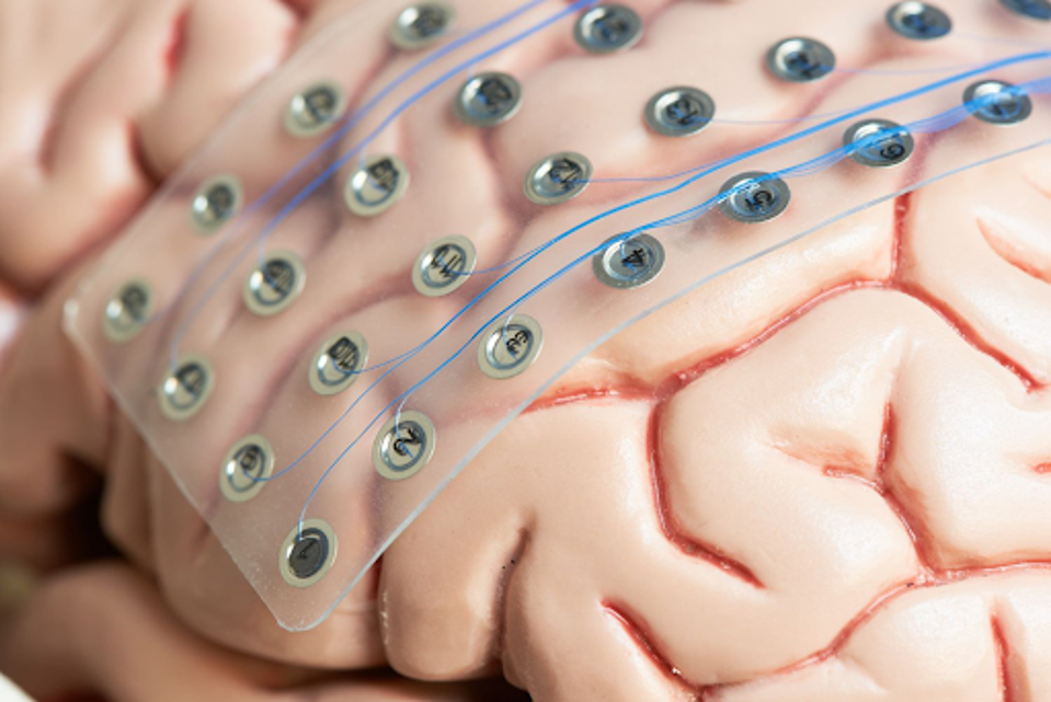
3D rendered model of an implanted electrode device for EEG monitoring.
GILLETTE CHILDREN’S
While implanted devices are perhaps the most accurate measurement avenue, they are expensive, difficult to maintain, and likely to cause brain damage.
Contrast implanted electrodes with scalp electrodes. These noninvasive alternatives are placed on the scalp surface, detecting brain cell activity through minuscule electrical charges. While perhaps less accurate than fully implanted electrodes, the process of applying the electrodes is much easier and less expensive.
Another measurement avenue of note is functional near-infrared spectroscopy. This non-invasive technique focuses not on the electrical signals themselves but on cortical hemodynamic activity. By detecting changes in cerebral blood flow, one can estimate neural patterns and activation of neurons in surrounding areas. While functional uses such as motor functions cannot be attempted with functional near-infrared spectroscopy, the method is an inexpensive and accessible alternative for brain physiology diagnostics.
Once the proper signals and waves are logged, there are a number of processing steps before they can be translated into data for the interface, namely filtering, feature extraction, and classification.
Filtering removes unwanted noise from the signal, whether environmental, involuntary motion by the patient or internal bodily reactions. Feature extraction highlights notable events in the recorded brain signal, for instance, oxygenation changes in hemoglobin when using functional near-infrared spectroscopy. Finally, classification is the labeling and sorting of signals and waves, including something as simple as right or left movement or as complicated as decoding thoughts for words.
As our grasp of brain-machine technologies continues to improve, so too will our understanding of how brain signals and waves work and can be used in these interfaces.
There are still a number of challenges that lay ahead when it comes to brain-machine interface research and development. Some specific challenges include the low signal-to-noise ratio of electroencephalography signals, prompting deciphering difficulties, as well as the non-stationary nature of signals and waves.
Our knowledge and understanding of brain signals, waves, and acquisition methods are crucial to the long-term success of brain-machine interfaces. I have no doubt that our brightest minds will continue to progress in this field that has the potential to revolutionize modern medicine as we know it.
Read Dr. Haseltine's latest piece with
![]()
