Is A Retinal Picture Worth a Thousand Diagnoses?
(Posted on Thursday, December 7, 2023)
This article was published on Forbes on 12/2/2023.
This story is part of a series on the current progression in Regenerative Medicine. This piece is part of a series dedicated to the eye and improvements in restoring vision.
In 1999, I defined regenerative medicine as the collection of interventions that restore to normal function tissues and organs that have been damaged by disease, injured by trauma, or worn by time. I include a full spectrum of chemical, gene, and protein-based medicines, cell-based therapies, and biomechanical interventions that achieve that goal.
The human eye is a marvelous work of art. Delicate as it is, it houses a complex system that enables us to see and experience the world around us. The core element of this intricate system lies within the retina – a delicate, light-sensitive tissue situated at the posterior part of the eye. Its crucial role is to convert incoming light into electrical signals, which are then elegantly processed by our brains, giving rise to our vivid perception of the world around us.
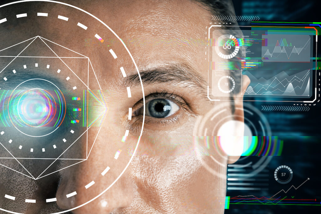
A healthy retina is paramount for crystal-clear eyesight. Any deviation from its optimal state can lead to an array of eye diseases, most of which can be diagnosed and tracked through retinal imaging. This type of imaging is a medical technology that enables ophthalmologists to capture and evaluate high-resolution, three-dimensional retina pictures, providing critical insight into a patient’s ocular health. Retina imaging technologies go beyond the traditional manual visual inspection with dilated pupils and instead offer a non-invasive and painless method to examine the retina, even when the patient is fully awake.
This imaging technology is essential in diagnosing and monitoring eye diseases such as diabetic retinopathy, age-related macular degeneration, and glaucoma. Even more surprising is that this technology can also lead to diagnoses of other conditions, such as neurodegenerative diseases like Alzheimer’s, Parkinson’s, and Huntington’s.
What is Retinal Imaging?
Retinal imaging is a non-invasive procedure revolutionizing the field of ophthalmology. It captures a digital image of the retina, optic disc, and blood vessels using a laser ophthalmoscope, providing a comprehensive view of the intricate structures within the eye. Unlike traditional ophthalmoscopy, retinal imaging offers a broader perspective, introducing more precision to eye exams and allowing for more accurate diagnoses.
One of the most notable breakthroughs in retinal imaging is Optical Coherence Tomography (OCT). Built upon light wave technology, OCT unveils the intricate layers of the retina with remarkable detail. By enabling ophthalmologists to examine the retina’s microscopic structures, this technology plays a crucial role in the early detection and diagnosis of various ocular conditions. The portability and cost-effectiveness of OCT are continually improving, ensuring that this invaluable tool is more accessible than ever before.
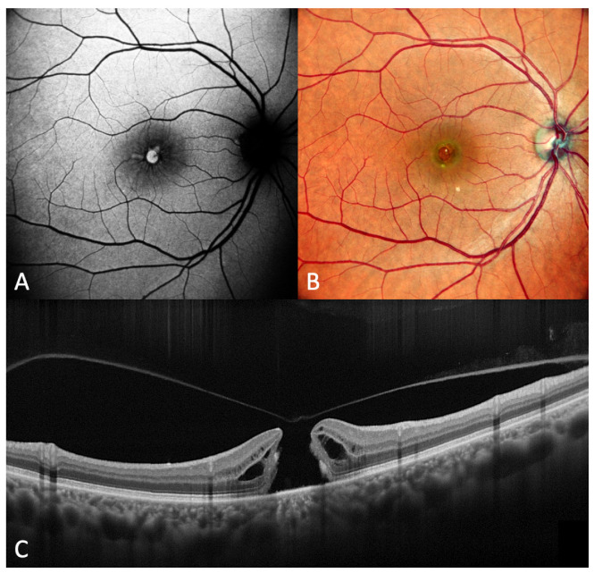
Compared to traditional retinal photography, OCT 3D retinal scanning offers a more comprehensive view of the retina’s structure, making detecting potential eye diseases at an early stage easier. This technology allows healthcare professionals to examine the retina beyond its surface, gaining a better understanding of its layers and structures. The process is painless, non-invasive, and takes less than five minutes to complete.
From Images to Diagnosis
Medical professionals can examine the retina non-invasively through retinal imaging and use qualitative and quantitative measurements to identify abnormal patterns that indicate various diseases.
A study by Fudan University in Shanghai explains how clinicians can use these images to diagnose different conditions. For instance, the thinning of the retinal nerve fiber layer, inner retinal layer, and choroidal layer, as well as reduced capillary density and abnormal vasodilatory response, have been linked to Alzheimer’s disease. These abnormalities can be seen with retinal imaging, allowing earlier diagnosis.
A comprehensive study from 2020 also showed promising results in using non-invasive polarimetric imaging of retinal amyloid deposits as a potential low-cost method for detecting Alzheimer’s disease. This technique involves using light waves to detect abnormal protein deposits in the retina, which have been linked to the development of Alzheimer’s disease.
In addition, a machine learning model has been developed using multimodal retinal imaging data to detect symptomatic Alzheimer’s disease. This model is based on the analysis of multiple imaging modalities, such as optical coherence tomography and fundus photography, to create a comprehensive picture of the retina and detect any abnormalities that may indicate the presence of Alzheimer’s disease.
These advances in retinal imaging technology offer a non-invasive and cost-effective approach to early detection of Alzheimer’s disease, potentially allowing for earlier intervention and improved patient outcomes.
Diagnosing More Than Eyes and Alzheimer’s
For example, in Parkinson’s disease, retinal changes such as thinning of the retinal nerve fiber layer and macular modifications can occur. Similarly, in Lewy body dementia, retinal thinning and reduced blood flow to the retina have been observed. In Huntington’s disease, retina degeneration can lead to vision deficits. In multiple sclerosis, a chronic autoimmune disease affecting the central nervous system, microcystic macular edema or outer retinal disruption can be potential biomarkers for early detection.
Early detection and intervention can lead to effective treatment and management strategies, potentially slowing the progression of these diseases and improving patients’ quality of life. With the help of retinal imaging, doctors can detect these diseases early, allowing for timely intervention and better outcomes.
Limitations of Retinal Imaging
Retinal imaging also has some limitations that are worth mentioning. One of the primary limitations of retinal imaging is that patients with cataracts or other media opacities may face challenges with the procedure. This is because these conditions can scatter light in the eye, interfering with the quality of the retinal image captured. Additionally, the cost of retinal imaging may be a barrier for some patients, although recent technological advances have made it more affordable.
It is also essential to note that retinal imaging is just one tool used for diagnosing and monitoring eye conditions. While it provides valuable information about the retina’s health, it cannot replace a thorough bedside examination performed by an experienced ophthalmologist. Therefore, retinal imaging should be used as a complementary tool to a comprehensive eye exam rather than a replacement for it.
To learn more about the eye, read more stories at www.williamhaseltine.com
Read Dr. Haseltine's latest piece with
![]()
