Wearable Ultrasounds: A Sonic Leap In Regenerative Medicine
(Posted on Thursday, December 7, 2023)
This article was originally published on Forbes on 12/7/2023.
This story is part of a series on the current progression in Regenerative Medicine. This piece discusses advances in wearable devices.
In 1999, I defined regenerative medicine as the collection of interventions that restore to normal function tissues and organs that have been damaged by disease, injured by trauma, or worn by time. I include a full spectrum of chemical, gene, and protein-based medicines, cell-based therapies, and biomechanical interventions that achieve that goal.
The ultrasound is a simple, yet versatile imaging system commonly used worldwide. Its most well-known application is in obstetrics and gynecology, but it can also be used for abdominal imaging, cardiology, and urology, among other fields.
Ultrasounds involve generating and transmitting sound waves through the patient’s body, encountering tissues, organs, and bones. These waves are returned to the ultrasound machine and processed to generate a real-time image. This is how the familiar ultrasound image of a fetus is developed, or in other cases, tumors, diseases, and so on.
However, conventional ultrasound probes are a dated technology, often susceptible to operator error and only resulting in a limited field of view. In a recent study for Nature Electronics, Dr. Lin Zhang and colleagues from the Massachusetts Institute of Technology developed a comfortable and flexible ultrasound patch. I will analyze their ultrasound patch and discuss its impact on long-term regenerative medicine.
The main draw of a flexible and conformable ultrasound patch alternative is to facilitate imaging on curved body parts, improve image quality, and enable prolonged monitoring. Until recently, there was difficulty selecting the right materials to build a smart flexible patch as ultrasound emitters require specific ceramics with specialized piezoelectric coefficients. With the recent advent of smart fabrics and stretchable technologies, the ingredients to develop the ultrasound patch were now available.
The bladder was selected as the focus for Zhang’s ultrasound patch due in part to the relative scarcity of wearable or portable ultrasound devices for abdomen monitoring, as well as the spatial requirements to measure the volume of the bladder in real-time.
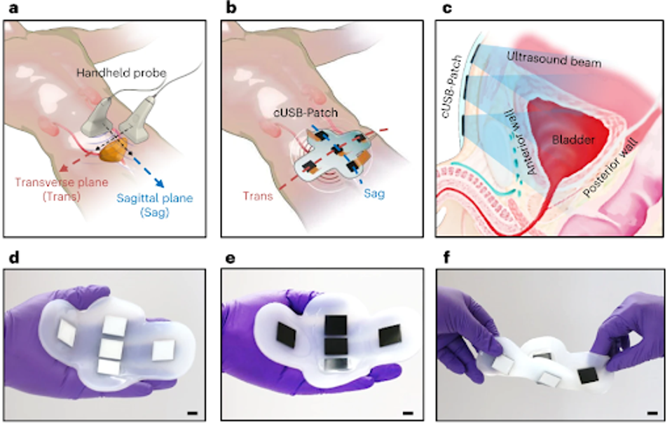
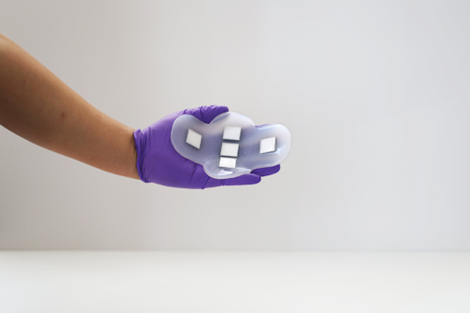
FIGURE 1: (A) Schematic of the operation of a handheld probe on the human’s lower abdomen for bladder imaging. (B) Schematic of the cUSB-Patch on the human’s lower abdomen for bladder imaging. (C) Schematic of a cross-sectional view of the cUSB-Patch on the lower abdomen. (D-F) The natural and flexible form factor of the cUSB-Patch: on hand with the view of the matching layer’s side (D), on hand with the view of the backing layer’s side (E) and the image of the cUSB-Patch under twisting (F).
The patch is made from a silicone rubber less than 5 mm thick, enabling significant stretchability and flexibility upon patient movement. As seen in the figure above, the five smart electrode patches emit a one-dimensional sound wave array that travels through the skin into the tissue of the targeted organ, in this case, the bladder.
The wave acquires a two-dimensional slice of the target organ, and that data is relayed back to the electrodes and a separate data bank. The flexibility of the patch allows for continued monitoring, even when the patient is in motion, removing the need for an office visit or medical administrator.
To evaluate their system, Zhang and colleagues conducted a human study to compare the performance of their ultrasound patch to a conventional ultrasound system. They analyzed the bladder volume of patients when they were full and partially empty, using the ultrasound systems to estimate volume both with and without ultrasonic gel, which is used to amplify and improve sound wave transmission.
Their investigations found that the ultrasound patch was very accurate in estimating the volume of the bladder in reference to the conventional ultrasound system. The ultrasound patch with and without gel could estimate bladder volume within the reasonable margin of error, whereas the conventional ultrasound without gel fell well short. These results suggest that the ultrasound patch is roughly as accurate as the gelled conventional ultrasound and much better than a gel-free conventional ultrasound.
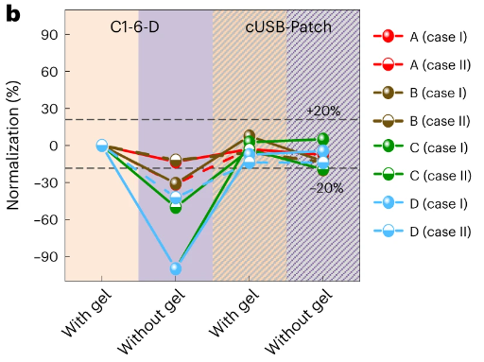
FIGURE 2: Normalization of the calculated bladder volumes of four subjects in eight different tests.
The applications for long-term therapeutic and regenerative medicine for the ultrasound patch are innumerable, only to be limited by the imagination of those implementing their use.
Among the most forthcoming are situations where someone may be unable to visit their physician for a medically-administered ultrasound. This could include working adults with children, elderly patients, or people with mobility limitations.
Another clear application is continual health monitoring for at-risk patients. As a wearable technology, patients may use the ultrasound patch in their daily lives without uncomfortability or cumbersomeness. This extends to disease monitoring and management for the already ill patient. Beyond urinary illness, ultrasounds may be used in cardiac settings, bone monitoring, and more.
Millions suffer from chronic illness that requires multiple repeat doctor visits on an annual basis. If the ultrasound patch can be adapted to reduce the difficulty on these patients and the load on our strained healthcare system, the ultrasound patch will soon be a resounding success.
Read Dr. Haseltine's latest piece with
![]()
